Scientific Summary Assessing prosthetic heart valves with CT
Scientific Summary
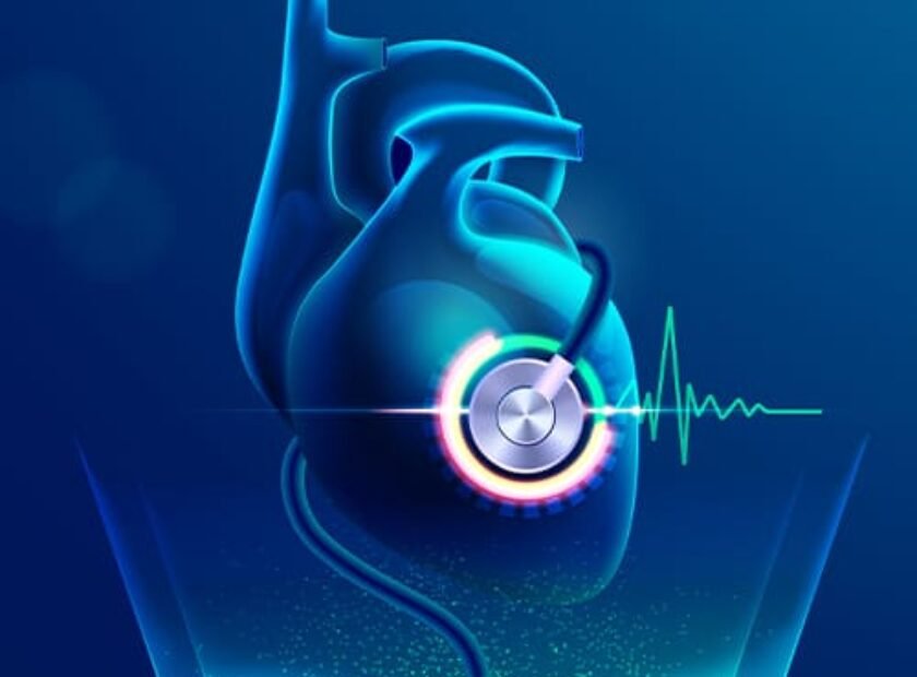
Assessing prosthetic heart valves with CT
Source: JACC. Jun 16, 2025.
A new multi-society consensus statement has been released to guide clinicians on the use of cardiac CT for evaluating prosthetic heart valves (PHVs). This document is the result of collaboration among major medical societies including RSNA, ACC, ESCV, SCCT, STS, and others.
Key Points:
1. CT vs. Echocardiography:
• While echocardiography remains the first-line imaging tool, cardiac CT offers complementary insights, especially in complex or ambiguous cases.
2. Types of Valves Covered:
• Includes mechanical and bioprosthetic valves used in:
• TAVR (Transcatheter Aortic Valve Replacement)
• SAVR (Surgical Aortic Valve Replacement)
• TMVR/SMVR (Transcatheter/Surgical Mitral Valve Replacement)
• Discusses both modern bileaflet designs and older models (e.g., tilting disc, ball-and-cage).
3. Topics Addressed:
• Imaging protocols and positioning
• Radiation and contrast considerations
• CT postprocessing techniques
• Assessment of structural valve deterioration (SVD) and dysfunction
Takeaway:
Cardiac CT is increasingly vital in evaluating prosthetic valve dysfunction. This consensus offers a structured approach to optimize filling a critical gap in existing practice.